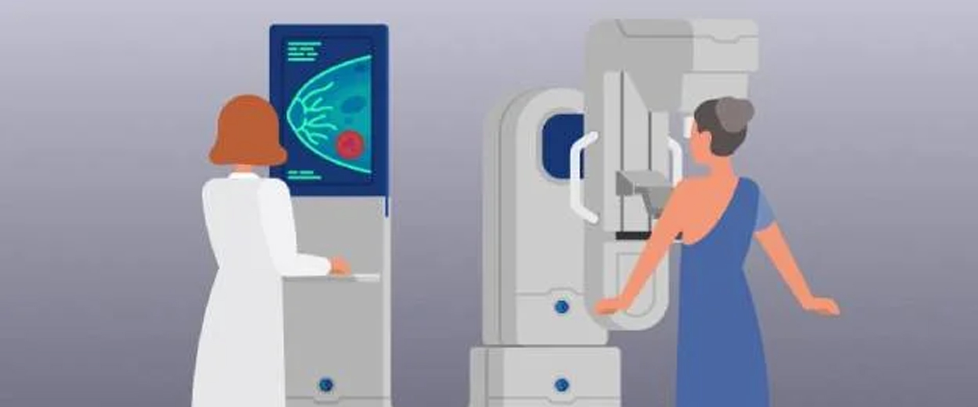
Mammography is a type of breast imaging that uses low-dose X-rays to detect early signs of breast cancer in women. Getting regular mammogram screenings is one of the most effective ways to detect breast cancer early when the chances of successful treatment and survival are highest.
Mammography plays a central part in early detection of breast cancers because it can show changes in the breast years before a patient or physician can feel them.
INDICATIONS FOR AMMOGRAPHY-
How does a Mammogram Work?
A mammogram is a quick and painless procedure where low-dose x-ray images of the breasts are obtained. During a screening mammogram, the breast is compressed between two plastic plates which spreads out the breast tissue to produce clear images. Two images are typically taken of each breast – one from top to bottom and another from side to side. The entire process takes around 30 minutes. The images are then inspected by a radiologist for any abnormalities such as masses, calcifications or other signs that may indicate breast cancer. At our diagnostic centre, Apex diagnostics, we do additional ultrasound of breasts in all the cases to co-relate the findings better.
Benefits of Regular Mammogram Screenings
Mammograms are proven to reduce the risk of dying from breast cancer. Getting regular screenings allows medical professionals to detect breast cancers early before they are large enough to feel or cause symptoms. According to studies, getting regular mammograms can reduce the risk of dying from breast cancer by 25-30% or more for women aged 40-74. When cancer is found early through a mammogram, treatment options are more effective and have a higher chance of cure.
HOW TO PREPARE FOR MAMMOGRAM -
Before scheduling a mammogram, the American Cancer Society (ACS) and other specialty organizations recommend that you discuss any new findings or problems in your breasts with your doctor. In addition, inform your doctor of any prior surgeries, hormone use, and family or personal history of breast cancer.
Do not schedule your mammogram for the week before your menstrual period if your breasts are usually tender during this time. The best time for a mammogram is one week following your period. Always inform your doctor or x-ray technologist if there is any possibility that you are pregnant.
How is the procedure performed?
Mammography is performed on an outpatient basis.
During mammography, a specially qualified radiologic technologist will position your breast in the mammography unit. Your breast will be placed on a special platform and compressed with a clear plastic paddle. The technologist will gradually compress your breast.
You must hold very still and may need to hold your breath for a few seconds while the technologist takes the x-ray.
When the examination is complete, additional ultrasound will be done for better co-relation fo your findings.
The examination process should take about 30 minutes.
What will you experience during and after the procedure?
You will feel pressure on your breast as it is squeezed by the compression paddle. Some women with sensitive breasts may experience discomfort. If this is the case, schedule the procedure when your breasts are least tender. Be sure to inform the technologist if pain occurs as compression is increased. If discomfort is significant, less compression will be used. Always remember compression allows better quality mammograms.
Who interprets the results and how do I get them?
A radiologist, a doctor trained to supervise and interpret radiology examinations, will analyze the images.
T our centre the average reporting time is about an hour and you will get your results on same day.
Limitations of Mammograms
No screening or imaging test detects 100% of breast cancers. There is a small chance of false positives requiring additional testing like ultrasounds or biopsies. Mammograms may not detect cancer in women with dense breasts where tumors are harder to distinguish from surrounding tissue. Additional screening methods like breast MRI may be recommended in such cases. It is important to discuss your breast density and family history with your doctor to determine the best screening option for you.
Factors That Increase Breast Cancer Risk
Certain factors increase a woman’s lifetime risk of developing breast cancer. Women with a family history of breast cancer in a parent, sibling or child have nearly twice the average risk and should have annual mammograms starting at an earlier age. Other high-risk factors include a personal history of breast cancer, exposure to radiation therapy to the chest before age 30 and certain genetic mutations like BRCA1/BRCA2.
Importance of Follow-up and Repeat Screenings
Mammograms may detect abnormalities that require further evaluation with additional tests like ultrasounds or biopsies. Any changes found even if initially thought to be non-cancerous still require close monitoring and repeat mammograms to check for progression or recurrence. Regularly following screening recommendations and promptly addressing any suspicious findings play a vital role both in early detection as well as long-term management of breast health and cancer prevention.
Conclusion
In summary, regular mammogram screenings remain one of the most reliable ways to detect breast cancer early and improve survival. Combining mammograms with monthly breast self-exams increases awareness of breast changes. Any discoveries should be promptly reported to your doctor. For women at high risk or with dense breasts, additional screening methods may be recommended. Let’s make mammography screening a priority to fight this disease. Book your consultation today!
Pre Insurance Health Check Partner.
We are committed to improving people’s lives through personalized health care. When you refer your patient to us, we are pleased to assist you with the diagnosis, treatment and monitoring of your patients’ care